Age-related macular degeneration (AMD)
Age-related macular degeneration (AMD) represents the first cause of deteriorating eyesight in patients in France over 65 years of age. By definition, AMD is a disease touching persons over 50 pages of age, affecting the central region of the retina (the macula), which is responsible for sharp central vision (reading, recognition of faces and details, driving a car…). The main recognized risk factors are age, heredity and tobacco. The Poitiers CHU participates in research protocols in the AMD area. Do not hesitate to contact us for further information. Several stages are commonly distinguished:
- The early stage:
Specialists do not yet speak of degeneration but rather of age-related maculopathy (ARM), which is characterized by drusen and/or alterations of the pigment epithelium. It can be responsible for moderate loss of vision or image deformation (metamorphopsia). Screening for this pathology necessitates regular surveillance of the fundus.
 |
 |
 |
| Age-related maculopathies | ||
- The more advanced stages:
- Dry or atrophic AMD is the most frequent and least severe form. It yields symptoms – loss of vision – only when the atrophic patches come together and touch the center of the macula.
- Humid or exuding AMD is related to proliferation of abnormal blood vessels from the choroid under the retina. Choroidian neo-vessels are fragile and can lead to a retinal edema or hemorrhages. Evolution is often rapid and without treatment, loss of central vision can occur in only a few weeks or months; that is the reason why emergency consultation and treatment are necessary.
 DMLA atrophique (patient 1)


 DMLA exsudative évoluée de l'oeil gauche (patient 1)


 |
 |
Treatment:
- For the atrophic form: At present, there exists no treatment able to halt its evolution, even though relevant research is making inroads. Treatment by vitamin supplements is aimed at slowing the evolution. Low vision rehabilitation and magnifying devices can be helpful with regard to the advanced forms of the disease. As a dry form can become humid, it is necessary to regularly monitor the fundus. Clinical studies concerning this pathology are ongoing.
- For the exuding forms: lntravitreous anti-VEGF injections currently represent the treatment of reference, and other molecules are likely to be marketed. The injections should be given on an emergency basis when the vision loss appears (humid AMD).
Knowledge and treatment of AMD are currently evolving due to recent advances in genetics, diagnosis (imagery) and therapy (anti-VEGF intravitreous injections). Highly active research offers hope for early treatment of the pathology and improved visual acuity in a significant number of cases. Clinical trails are ongoing. Do not hesitate to contact us for further information.
Retinal detachment

Retinal detachment by macular hole in a case of high myopia This is a severe, often rapidly appearing pathology, and it necessitates surgical treatment. It consists in detachment from its support (the pigmentary epithelium) of part of the retina surface, with liquid being interposed between the two layers. It is most often secondary to a retinal tear. The main risk factors are myopia, posterior detachment of the vitreous body, past history of retinal detachment in the other eye, and past history of cataract surgery. Symptoms : Visual field amputation followed by complete loss of vision if the retinal detachment becomes total. It is at times preceded by sensations of peripheral flashing lights (phosphenes), which translate traction on the retina, or a form of ” black soot” invading the view, which translates a small hemorrhage due to the retinal tear.

Treatment : 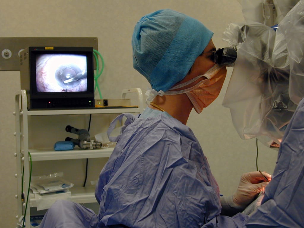 At the “tearing” stage, preventive treatment by local laser application reduces the risk of evolution towards retinal detachment. At the detached retina stage, it is necessary to undergo surgery; left untreated, retinal detachment leads to blindness. Treatment of a detached retina is surgical alone. The applied techniques vary according to the source of the detachment and the condition of the vitreous body. After the operation, rest and close monitoring are necessary, and perhaps specific positioning. Ambulatory treatment is possible.
The epiretinal membranes and macular holes
Membranes can form at the surface of the macular retina, the macula being the central part of the retina. They frequently occur in the general population, and surgical treatment is necessary only when visual acuity is particularly altered or when bothersome deformities appear. The operation takes place under simple local anesthesia by means of transconjunctival micro-incisions.
 |
 |
 |
 |
 |
 |
Diabetic retinopathy

This is a complication of a diabetes characterized by lesions affecting the retinal capillaries and venules (small vessels), which can be blocked, leading to retinal suffering. Abnormal vessels (neo-vessels) then proliferate on the retinal surface, possibly leading to complications (severe stage of the disease). Diabetic retinopathy appears after several years of diabetes evolution. Unbalanced diabetes over several years constitutes the main risk factor; if it is not mastered, it can lead to loss of the sense of vision. At the onset of diabetic retinopathy, no symptom is felt by the patient. Vision is conserved. Screening by means of yearly eye fundus is consequently necessary. Even the advanced forms of diabetic retinopathy may not involve symptoms; that is why regular screening by an ophthalmologist is so vitally important. At an evolved stage, a sudden or progressive loss of vision attests to the appearance of serious complications such as macular edema, which reduces central vision, intravitreal hemorrhage or detached retina. Aspect of diabetic retinopathy with numerous retinal hemorrhages and a macular edema surrounded by exudates.

Eye fundus imaging is the main screening exam. At the early stage, diabetic retinopathy necessitates no specific treatment. Strict diabetes control and control of other cardiovascular risk factors such as arterial hypertension are indispensable to limitation of diabetes progression. At the evolved stage, retinopathy treatment essentially consists in a series of laser sessions. Laser treatment is aimed at destroying the ischemic areas of the peripheral retina so as to avoid serious complications. In the event of macular edema, intraocular injections of corticoids or anti-angiogenic drugs is necessary.
| Proliferative diabetic retinopathy before surgery | Proliferative diabetic retinopathy after surgery (same patient) |
 |
 |
| Proliferative diabetic retinopathy of the right eye before and after surgery (angiography). | |
 |
 |
|
Proliferatuve diabetic retinopathy of the left eye before and after surgery (angiography). |
|


Retinography for diabetics 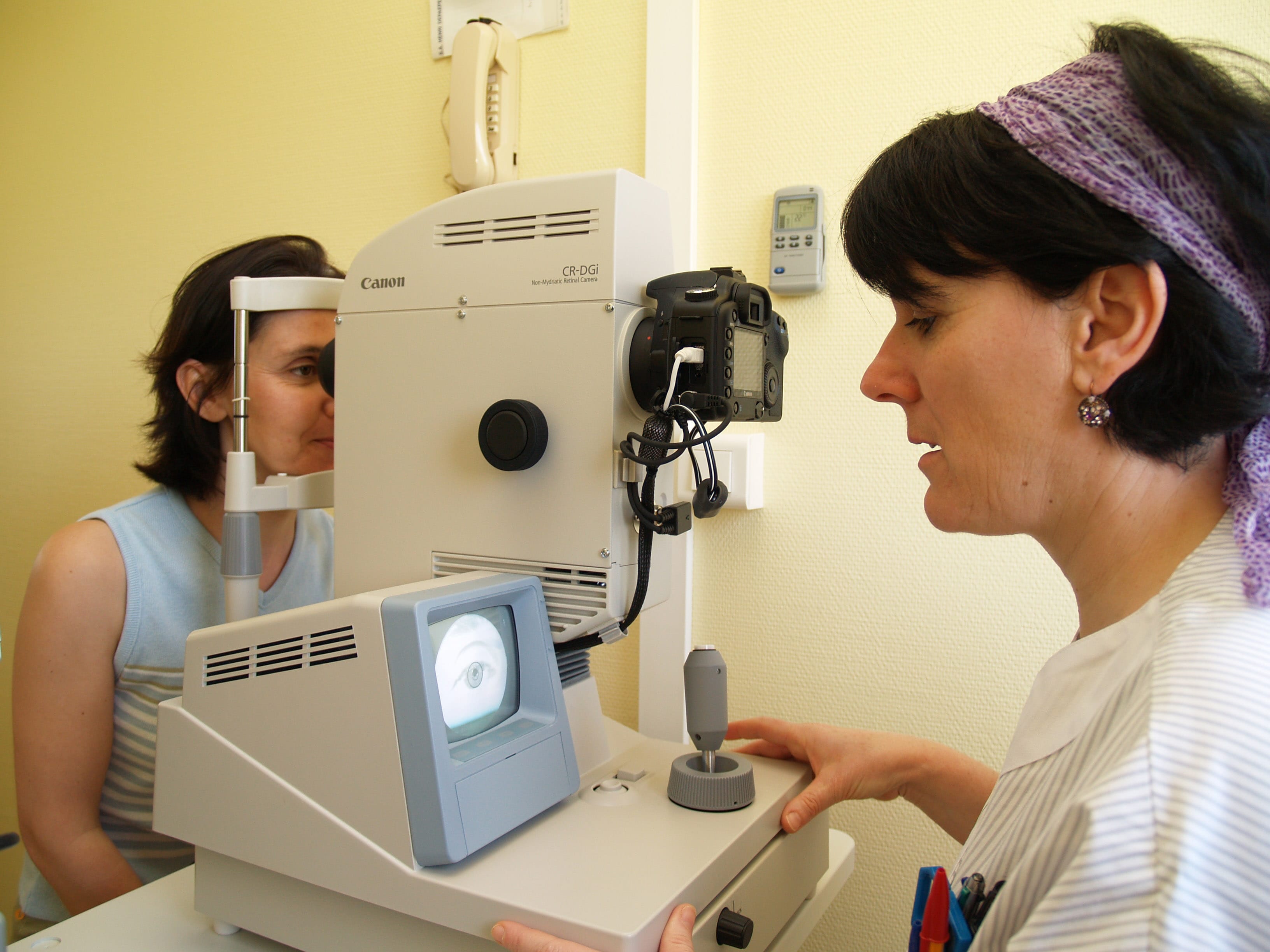 A non-mydriatic retinograph has been put into place in the departments of endocrinology and ophthalmology. It is used for screening of diabetic retinopathy without recourse to pupil dilation, and is an alternative to the funduscopic examination.It ensures improved monitoring of diabetic patients subject to retinal complications, and who must be examined one a year in view of detecting, as early as possible, any problem justifying specialized treatment.The digital photos taken by a nurse during the diabetology consultation are interpreted in the ophthalmology department by a trained expert. As a result, the patient is directly treated in a single department. Using this technique, it is possible to analyze the photos of 60 diabetic patients a week and thereby better meet existing demand. Depending on his retinal condition, a patient can either be summoned to the ophthalmology ward or referred back to his usual ophthalmologist. To sum up, non-mydriatic retinography improves screening of the retinal complications of diabetic patients, thereby carrying out good practice recommendations and rationalizing ophthalmology consultations as patients with patent retinopathy are given privileged access to ophthalmologists. A non-mydriatic retinograph has been put into place in the departments of endocrinology and ophthalmology. It is used for screening of diabetic retinopathy without recourse to pupil dilation, and is an alternative to the funduscopic examination.It ensures improved monitoring of diabetic patients subject to retinal complications, and who must be examined one a year in view of detecting, as early as possible, any problem justifying specialized treatment.The digital photos taken by a nurse during the diabetology consultation are interpreted in the ophthalmology department by a trained expert. As a result, the patient is directly treated in a single department. Using this technique, it is possible to analyze the photos of 60 diabetic patients a week and thereby better meet existing demand. Depending on his retinal condition, a patient can either be summoned to the ophthalmology ward or referred back to his usual ophthalmologist. To sum up, non-mydriatic retinography improves screening of the retinal complications of diabetic patients, thereby carrying out good practice recommendations and rationalizing ophthalmology consultations as patients with patent retinopathy are given privileged access to ophthalmologists. |